


01 · The Patient
A 66-year-old man with type II diabetes of approximately twenty years’ duration was referred to our unit on June 10, 2025, after three years of recurrent ulcerative episodes involving the left foot and lower limb.
The most recent lesion had been present for about two years and was associated with extreme pain. His clinical history was highly complex: cardiac insufficiency due to hypokinetic-dilated cardiomyopathy, a dual-chamber ICD, previous mitral valve repair, chronic venous insufficiency with previous right gemellar deep vein thrombosis, iatrogenic hypothyroidism and diabetic peripheral artery disease.
His pharmacological therapy included pantoprazole, bisoprolol, dapagliflozin, terazosin, valsartan, canrenone, amiodarone, cilostazol and warfarin, in addition to peripheral and central analgesics without a scheduled regimen.
Before referral, he had been followed by non-specialist practitioners and had undergone a wide range of local treatments without meaningful clinical benefit.
02 · The Wound
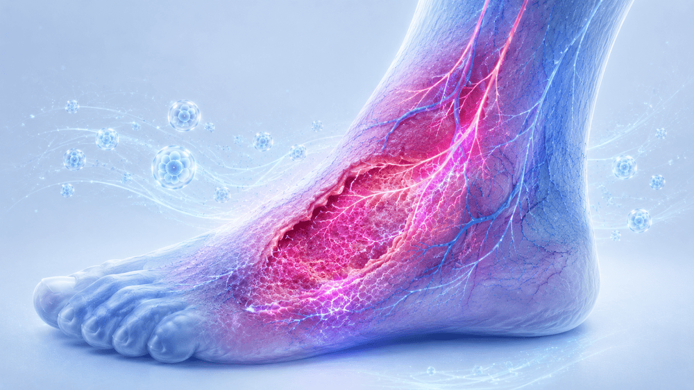
At presentation on June 10, 2025, the wound involved the left foot and lower limb. The lesion measured 19.13 cm² and had not healed for approximately two years.
Pain was very severe, with an NRS score of 9. This made local management, compression and mobility extremely difficult.
The wound was part of a multifactorial diabetic foot condition, in which neuropathic, vascular and systemic cardiac factors overlapped. The reduced perfusion reserve caused by heart failure created a particularly unfavorable biological environment for wound repair.
03 · Prior treatment history
After clinical examination, arterial Doppler ultrasound showed preserved distal vascularization through the three axes, but reduced flow in the anterior tibial arteries.
Given the cardiac pump deficit and the relevance of the angiosome concept, a percutaneous transluminal angioplasty of the left anterior tibial artery was performed on June 28, 2025. The procedure restored flow successfully and had no immediate postoperative complications.
However, at follow-up on July 8, 2025, a revascularization syndrome was observed, characterized by marked edema and elevated inflammatory markers. This progressed to erysipelas despite immediate antibiotic therapy, and the subsequent phase was further complicated by heart failure and hospitalization.
On July 30, 2025, the patient returned with an enlarged lesion measuring 21.64 cm² and persistent uncontrolled pain.
04 · Decision and protocol
After local treatment with a topical antibiotic combined with hyaluronic acid gel to prepare the wound bed, mononuclear cell therapy was indicated, subject to approval from the cardiology team.
Given the successful revascularization but the patient’s persistent biological fragility, the decision was made to administer advanced PRP in combination with CGF using a staged approach.
The treatment was performed with the High Q Cell® All-In-One procedural kit, which allows the preparation of both E-PRP and CGF from autologous blood.
The rationale was to combine injectable biological stimulation with a local fibrin matrix, aiming to improve perfusion, reduce inflammation and support progression toward tissue repair in a complex diabetic and cardiopathic patient.
05 · Clinical response
On September 15, 2025, the patient underwent the first E-PRP and CGF grafting procedure. The procedure was uneventful.
On October 30, 2025, the wound had improved and pain management was better, with the NRS score decreasing to 4 following analgesic therapy. A second E-PRP and CGF graft was performed as scheduled, again without complications.
Because the patient also had chronic venous insufficiency related to a previous deep vein thrombosis, edema control remained a major component of management. As pain became more manageable, short-stretch elastic compression could be initiated, which had previously been impossible.
By December 3, 2025, the lesion had significantly reduced to 7.59 cm². However, pain had not improved further, so an additional cycle of two treatments with E-PRP and bioengineered grafting was planned with the patient’s consent and cardiology approval.
06 · Outcome and follow-up
On December 15, 2025, the first treatment of the additional cycle was performed without postoperative complications. After this session, lesion-related pain decreased to NRS 2.
Encouraged by this response, the treatment cycle was completed as planned. On January 24, 2026, the final PRP and CGF application was performed.
After the final application, pain disappeared completely, reaching NRS 0. Analgesic therapy could be discontinued, and the patient’s general and cardiac condition progressively improved.
On February 25, 2026, the lesion had drastically reduced to 2.24 cm² with no pain. On March 17, 2026, the wound was beginning to close, with an area of only 1.17 cm² and persistent absence of pain.
Overall, the target lesion achieved a 90% area reduction over six months, from 11.4 cm² to 1.17 cm². Pain dropped to NRS 0 after the third application and remained controlled. The patient resumed normal motor activities, with improvement in his general and cardiac condition.
07 · Discussion
This case illustrates the role of E-PRP and CGF as part of a multidisciplinary strategy in a diabetic patient with severe cardiovascular comorbidity.
The clinical challenge was not limited to the wound itself. The lesion existed within a complex balance of diabetic arteriopathy, impaired cardiac pump function, venous insufficiency, edema, revascularization syndrome, erysipelas and hospitalization for heart failure.
In this context, the minimally invasive nature of PBMNC therapy was particularly relevant. It allowed treatment to be tailored to a fragile patient in whom aggressive options carried significant risk.
The response suggests a synergistic biological effect: improved local perfusion, reduction of pro-inflammatory factors, and support for the transition of the wound bed toward a more anabolic and reparative phase.
The most clinically meaningful outcomes were the disappearance of pain, discontinuation of analgesic therapy, recovery of mobility and substantial wound reduction. These results support the potential role of E-PRP and CGF as adjunctive regenerative tools in selected complex diabetic foot cases, especially when cardiovascular comorbidities limit the therapeutic margin.