


01 · The Patient
An 80-year-old woman with a long history of multiple recurrent ulcers of the lower limbs was followed at our center after several previous episodes of ulceration.
The patient was slightly obese, with a BMI of 33.6, and her clinical history was marked by several comorbidities. She had diabetes mellitus, hepatitis C acquired during a previous hospitalization, hypertension with hypertensive cardiopathy, chronic atrial fibrillation under anticoagulant therapy, and depressive syndrome.
Her surgical history included tonsillectomy, hysterosalpingectomy, thyroidectomy for goiter and left mastectomy for cancer three years before presentation.
Pharmacological therapy included letrozole, levothyroxine sodium, pantoprazole, sotalol, atorvastatin-perindopril lysine-amlodipine, metformin and rivaroxaban.
The case was particularly complex because the patient had already received previous diagnoses and treatments, yet the recurrence pattern suggested that the clinical picture needed to be reassessed.
02 · The Wound
On July 1, 2025, the patient presented with multiple spontaneous recurrent ulcers on both lower limbs.
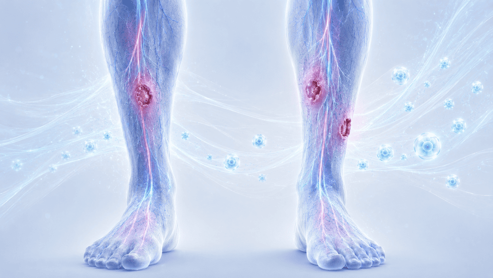
The lesions involved the anterior aspect of the left leg, the anterior aspect of the right leg and the lateral aspect of the right leg. The total lesion area measured 15.32 cm² and the wounds were painful, with an NRS score of 7.
The lesions had initially been interpreted in the context of a previous vasculitis diagnosis. However, the clinical behavior of the wounds, their recurrence and the intensity of pain raised doubts about the completeness of the previous diagnostic framework.
The wounds were not behaving as expected under the previously adopted therapeutic approach, making a full diagnostic reassessment necessary.
03 · Prior treatment history
The patient had been known to our center since 2023 for multiple leg ulcers.
In March 2023, blisters appeared on the lower limbs and were diagnosed as erysipelas. They were treated with zinc-coated dressings. A Doppler ultrasound revealed unspecified stenosis of the tibial arteries. Lipoid necrobiosis was diagnosed and resolved after four weeks.
In October 2023, a recurrence occurred. After a Doppler ultrasound showed a stable vascular picture, debridement and engineered skin grafting were performed, leading to resolution in September 2024.
When the patient returned in July 2025 with a new recurrence, the previous treatment strategy was resumed. However, despite steroid therapy and an engineered graft, the lesions did not improve.
This lack of response suggested that the active driver of the wounds had not been fully identified.
04 · Decision and protocol
In September 2025, bilateral transcutaneous oximetry was performed. The results were strongly suggestive of a vascular problem, with saturation values of 11 at the left ankle and 14 at the right ankle.
A repeat Doppler ultrasound was therefore requested. This revealed stenosis of the common femoral artery with occlusion of the tibiofibular trunk in the right leg, while the left leg showed pre-occlusive stenosis of the popliteal artery.
The patient was referred for angioplasty. However, after returning from surgery, the lesions worsened significantly due to revascularization syndrome.
At that point, the patient was placed on the waiting list for peripheral blood mononuclear cell transplantation.
The treatment strategy combined E-PRP, to promote vascular regeneration and modulate inflammation, with coverage of the lesions using a porcine dermal graft.
05 · Clinical response
On October 13, 2025, the patient was admitted with severe pain, with an NRS score of 8. The total lesion area had increased to 38.5 cm² as a consequence of the revascularization syndrome.
After preparation, a mononuclear cell graft was performed using the High Q Cell® Liquid procedural kit. The lesions were then covered with a porcine dermal graft. The postoperative course was uneventful.
On November 27, 2025, the treatment was repeated, again without significant complications.
The wounds began to respond clearly. Pain became fully controlled and decreased to a well-tolerated discomfort. The patient spontaneously discontinued analgesic medication.
At the January 13, 2026 follow-up, the total lesion area had already decreased to 5.56 cm², with pain reduced to NRS 1.
06 · Outcome and follow-up
On March 17, 2026, five months after the beginning of treatment, the results were highly significant, although the patient remained under evaluation.
The lesion on the anterior aspect of the left lower limb measured only 0.81 cm². The anterior lesion on the right lower limb had resolved. The lateral lesion on the right lower limb had decreased to 0.37 cm².
Pain was virtually absent, with an NRS score of 1, and analgesic therapy was permanently discontinued.
Transcutaneous oxygen saturation reached 45% bilaterally. Diabetes, which had previously been poorly controlled, finally showed glycated hemoglobin values within the normal range.
The patient regained complete independence and was able to return home with limited assistance from her children.
07 · Discussion
This case illustrates the importance of reassessing the entire diagnostic picture when a wound does not behave as expected.
The patient had multiple bilateral recurrent ulcers and had previously been managed according to an inflammatory or autoimmune hypothesis. However, the persistence of uncontrolled, piercing pain suggested that the vascular component had been underestimated.
The decisive test was oximetry. Its results revealed a severe vascular problem that had not been fully recognized, and this was later confirmed by Doppler ultrasound.
After angioplasty, the patient developed revascularization syndrome, with rapid worsening of the lesions. In this context, E-PRP was selected to support the vascular recovery phase and modulate the intense inflammatory response.
The treatment appeared to act on two key components: the vascular impairment, by supporting collateral vessel formation and oxygen supply, and the pro-inflammatory state, by promoting the transition from inflammatory catabolism toward reparative anabolism.
The clinical outcomes were meaningful: TcPO₂ increased nearly fivefold, lesion area decreased by approximately 85% at 12 weeks after treatment, pain was dramatically reduced and glycemic control normalized.
The case confirms that complex chronic ulcers should not be interpreted only through a previous diagnosis when the clinical course changes. A full reassessment can reveal hidden drivers of non-healing and open the way to more targeted regenerative treatment.