


01 · The Patient
An 80-year-old woman was referred to our care in July 2024 with severe pain in her left big toe, associated with an apical phlycten and clear signs of acral vascular compromise.
Her medical history primarily included scleroderma with Raynaud’s disease. The clinical picture was further complicated by hypertension and chronic atrial fibrillation, for which she was treated with direct-acting oral anticoagulants. She also tested positive for Lupus Anticoagulant, increasing her thrombotic risk.
From a vulnological standpoint, the patient had already undergone amputation of the first to third toes of the right foot in May 2023. She had also undergone percutaneous transluminal angioplasty of the right femoral artery, and a Doppler ultrasound performed in April 2024 had revealed bilateral chronic peripheral arterial occlusive disease.
Her pharmacological therapy included dabigatran etexilate, olmesartan medoxomil with hydrochlorothiazide, allopurinol, aminaftone, digoxin, atenolol, colchicine, paracetamol and pregabalin.
02 · The Wound
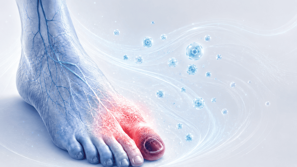
At presentation on July 9, 2024, the left big toe was cyanotic and painful, with an apical blister. The clinical appearance was consistent with Raynaud’s disease complicated by acral ischemia.
The lesion area measured 7.59 cm² and pain was severe, with an NRS score of 9.
The wound was not a standard ulcerative lesion. It developed in the context of acral vasoconstriction, ischemic pain, systemic autoimmune disease and bilateral peripheral arterial disease. The previous contralateral toe amputations made the preservation of the left big toe particularly important for ambulation and quality of life.
03 · Prior treatment history
At first evaluation, vasodilator therapy was initiated in consultation with the rheumatology department.
Further assessment was performed using Doppler ultrasound, which fortunately ruled out the need for revascularization. This confirmed that the patient’s immediate management had to focus on controlling the underlying disease and supporting local tissue survival.
Despite standard treatment, the local condition worsened. On December 12, 2024, the patient returned with progression of the lesion and initial acral ischemia. The lesion area had decreased to 1.73 cm², but pain remained severe, with an NRS score of 8, and the clinical risk of amputation had become evident.
Given the previous contralateral amputations and the worsening condition, an alternative limb- and toe-salvage strategy was considered necessary.
04 · Decision and protocol
Because of the risk of amputation and the patient’s previous loss of toes on the contralateral foot, treatment with E-PRP and CGF was proposed.
A triple application cycle was selected in an attempt to save the big toe and forefoot.
The first procedure was performed on December 19, 2024, using the High Q Cell® All-In-One procedural kit. E-PRP and CGF were obtained from autologous blood during the same procedure.
The biological rationale was to use E-PRP to support collateral circulation and reduce local inflammation, while CGF provided a dense fibrin matrix capable of sustaining local regenerative activity over time.
05 · Clinical response
On January 13, 2025, follow-up showed excellent recovery, with restoration of vascularization at the base of the first phalanx and only a minimal area of acral ecchymosis.
On February 11, 2025, at preoperative check-up, the lesion measured 1.12 cm². Pain had decreased but remained intense, with an NRS score of 5.
The second E-PRP graft was then performed under analgesic sedation. The injection targeted the angiosome of the anterior tibial artery and the first metatarsal area of the foot, and was combined with CGF application using the same procedural kit. The postoperative course was uneventful.
On February 18, 2025, the patient was discharged with optimal recovery, a warm foot and decreasing pain, with an NRS score of 4.
06 · Outcome and follow-up
On March 4, 2025, the third and final E-PRP injection graft was performed in the anterior tibial angiosome area and in the interosseous spaces of the metatarsals throughout the forefoot, together with CGF grafting. The postoperative course was uneventful.
From this point onward, the lesion entered the resolution phase. Pain remained controlled, and treatment continued with advanced local hydrofiber dressings until closure.
On April 7, 2025, the lesion measured 0.55 cm², with pain still decreasing at NRS 4. On May 20, 2025, the lesion had further reduced to 0.34 cm², while pain had dropped to NRS 2.
On July 22, 2025, the lesion measured 0.24 cm² and pain had become only slight discomfort, with an NRS score of 1. On October 14, 2025, the lesion area remained stable at 0.24 cm², but it was clearly healing and pain had completely disappeared, reaching NRS 0.
By December 19, 2025, ten months after the first E-PRP and CGF treatment, complete resolution was achieved. At the last follow-up visit in May 2026, the patient remained disease-free.
07 · Discussion
This case illustrates the possible role of PBMNC therapy in Raynaud’s syndrome complicated by peripheral arterial disease and acral ischemic progression.
The case was clinically significant because standard vasodilator therapy alone was insufficient to prevent worsening. The patient had already undergone contralateral toe amputations, so preserving the left big toe was essential for balance, ambulation and quality of life.
The response was progressive and linear, which is particularly important in Raynaud-related ischemic lesions. In this condition, long healing times are not necessarily a negative indicator; what matters is disease control, stabilization of tissue viability and consistent reduction of pain and lesion size.
The three E-PRP and CGF applications appeared to support local vascular recovery, reduce ischemic pain and help prevent progression toward amputation.
This treatment should not be interpreted as an alternative to the standard management of Raynaud’s syndrome or peripheral arterial disease. Rather, it may be considered as part of an appropriate treatment plan in selected cases where acral ischemia progresses despite standard therapies and tissue preservation is a priority.